The structure of the urinary system and its diseases
The human body is a “full-cycle factory”, constantly producing many substances, many of which are harmful and must be eliminated from the body. There are several ways to do this. All harmful substances are excreted with breath, sweat, feces and urine. Thus, the urinary system is one of the main ways to get rid of everything harmful and unnecessary to the body. Its structure and diseases will be discussed today.
The most important organ that plays a leading role in detoxification processes. It is paired, but it is possible to exist with one, and with genetic disorders, the kidney can be doubled. They are parenchymal organs. Located in the lumbar region. The structure of the body is quite complex. The organ consists of:
- Capsules and barks. The nephrons are immersed in it, in which the primary urine is formed. A glomerulus of capillaries is hidden in the nephron, which is needed to filter water, urea and layers.
- medulla. Primary urine passes through its tubules. They also carry out the return of glucose and the remaining water to the capillaries. After that, secondary urine remains, which enters the pyramids of the kidney.
- renal pelvis. Secondary urine enters it from the pyramids and is sent to the ureters.
- Kidney gate. Here, an artery enters the organ and a vein exits. They are also the entrance to the ureters.
- Inside the organ are: renal column, adipose tissue, papilla, renal sinus and calyces (small and large).
The normal weight of the kidney is about 200 g, the thickness is about 4 cm, the length is from 10 cm to 12. If the right kidney is slightly lower than the left, this is normal.
The main functions of the urinary system are:
- getting rid of unnecessary and waste products of metabolism;
- maintenance of homeostasis (meaning the water-salt balance);
- hormonal function (carried out by the adrenal glands).
Several organs work for all this at once:
- kidneys;
- ureters;
- bladder;
- urethra.
There are also secondary, but no less important organs, such as the aorta and the inferior vena cava, as well as the adrenal glands, which are glands that synthesize hormones, including adrenaline and norepinephrine.
Ureters
They are thin and long tubes extending from the pelvis and flowing into the bladder. The ureters connect the bladder and pelvis. The walls of the organ consist of mucous (stratified epithelium), muscular and adventitious (connective tissues) layers. They are located in the retroperitoneal space, have a length of 28 - 34 cm, but the left one is usually slightly longer, due to the location of the kidneys. The basis of the organ is smooth muscles, the outer layer is connective tissue, and the epithelium inside. It has the ability to peristalsis, in the region of the mouth, in the middle of the organ and in the region of connection with the pelvis, it has constrictions.
Bladder
A fairly large organ located in the pelvis. It is an organ of smooth muscle, which is lined with epithelium inside. From above it is covered with a peritoneum. Comprises:
- necks;
- side, back and front walls;
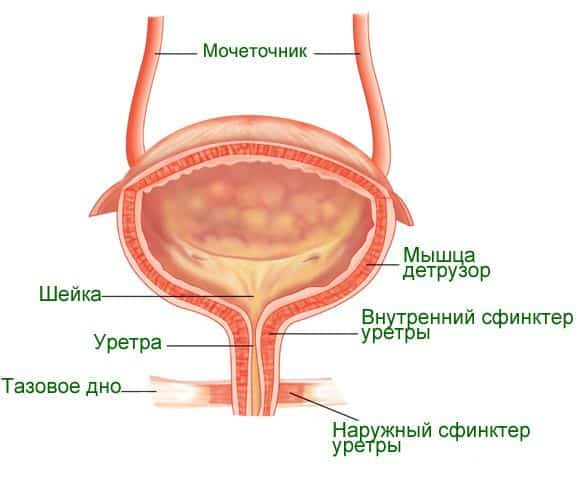
The mouths of the ureters are located on the back wall of the organ. It has the form of a bag, reaching a volume of 200 - 400 ml when filled. Urine accumulates for about three hours, when the walls contract, it leaves the urethra.
Urethra
Also called the urethra. In women and men, this organ has differences in structure:
- It is a tubular and unpaired organ.
- Consists of smooth muscle, which is lined from the inside with epithelial tissue. Its task is to bring urine into the external environment. Like the ureters, it has three layers. In men, it is also needed for ejaculation and is located in the penis. The female urethra is wider, well stretched, slightly shorter and easily affected by infection.
Diseases of the urinary system
Unfortunately, all organs of the urinary system are prone to disease. Here are the most common ailments of this organ system.
Bladder:
- hyperactive;
- neurogenic;
- (including interstitial);
- hernia;
- diverticulum;
- Marion's disease;
- tumors and cancer;
- sclerosis of the neck of the bladder;
- stenosis of the neck of the bladder;
- structural anomalies.
Ureters:
- strictures;
- stones in the ureters;
- Ormond's disease;
- reflux vesicoureteral;
- ureterocele;
- neuromuscular dysplasia;
- empyema of the stump of the organ;
- tuberculosis of the ureters;
- tumors.
Kidneys:
- structural anomalies;
- pyelonephritis chronic and acute;
- cyst;
- nephroptosis (omission);
- glomerulonephritis;
- hydronephrosis;
- jade apostematous;
- paranephritis;
- abscess;
- pyonephrosis;
- carbuncle;
- nephropathy (diabetic, during pregnancy);
- acute and chronic renal failure;
- tumors;
- tuberculosis;
- syndrome of prolonged compression of the kidney.
Urethra:
- fistulas;
- urethritis;
- anomalies (congenital narrowing, doubling, epispadias, hypospadias);
- stricture;
- prolapse (including the mucous membrane);
- diverticulum;
- papillomas (they are condylomas);
- polyps;
- angioma;
- fibroma;
- caruncle;
- trauma;
- tumors are malignant.
To diagnose any ailments of the urinary system, examinations such as laboratory diagnostics (urine and blood tests), cystoscopy, x-ray methods, ultrasound, MRI, CT should be carried out. Symptoms can vary widely, but with many urinary tract disorders, urination disorders, pain, and changes in the appearance of urine can be noted.
The urinary system is one of the largest organ systems in our body. Its main task is to free the body from toxins. Not only the kidneys work for this, but also the ureters, bladder and urethra.
You can also learn about the urinary system in this video.