Urinary system. Anatomical and physiological features of the urinary system
Isolation is the process of removing the end products formed as a result of metabolism from the body. The organs involved in excretion include the intestines, lungs, and skin. However, more than 90% of the compounds are excreted by the urinary system. Let's take a closer look at its structure.
The structure and functions of the urinary system
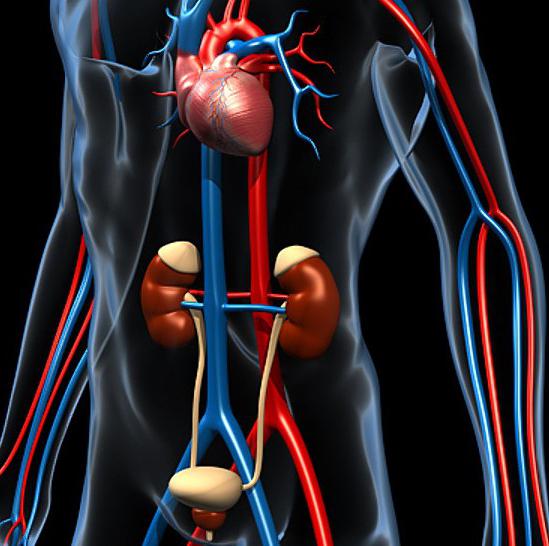
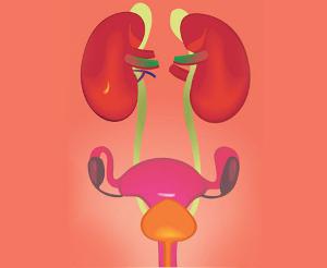
Several organs are involved in the excretion process. These include the kidneys, ureters, urethra, and bladder. In some organs, metabolic products are processed. By means of others, in fact, the withdrawal is carried out. So, the formation of urine occurs in the kidneys, the accumulation - in the bladder. Excretion is carried out through the urethra. The kidneys are a paired organ. The system also contains the ureters. They are presented in the form of hollow tubes. With their help, the kidneys and the urinary system, presented in the form of the urethra and bladder, are connected. The latter is a hollow organ. It is located in the small pelvis. The connection of the bladder with the surface of the body is carried out through the urethra. Unlike other organs, this channel has differences depending on gender. This is the general structure of the human urinary system.
Filtration organs
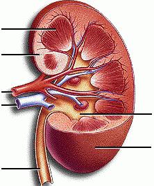
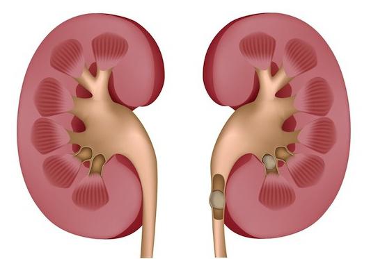
On the sides of the spinal column, in the area between the 12th thoracic and 2nd lumbar vertebrae, are the kidneys. They are adjacent to the back of the abdominal cavity. The right kidney is slightly lower than the left. The organs are bean-shaped. In the kidneys, the posterior and anterior surfaces, the lower and upper ends, as well as the medial and lateral edges, are distinguished. On the concave area (medial), facing the spine, there are gates. They include arteries and nerves, lymphatic vessels, a vein, and an ureter exit. The fixation of the kidneys is facilitated by the membrane that covers them. Fibrous tissue is adjacent to the substance of the organ. Outside of this shell is a living capsule. Front and back it is surrounded by the fascia of the kidney. In front, the organ is covered by the peritoneum. Strengthen the fixation of the kidneys intra-abdominal pressure and blood vessels. They enter and exit the body. The kidney contains a cortex. Its thickness is 5-7 mm. It is located on the periphery. The medulla is also present in the kidney. It consists of 7-12 pyramids. The medulla is turned to the cortical base, and to the renal sinus - by the apex. There are also pillars in the organ. They are formed due to the cortical substance, wedged into the medulla.
Structural units
They are nephrons. This is a system of renal tubules involved in the formation of urine. One nephron can have a length in the range of 18-50 mm, while their total length is about 100 km. The number of elements in each kidney is about a million. The nephron includes a capsule and a three-linked tube. It, in turn, consists of the proximal part of the convoluted tubule of the first order, the loop and the distal part of the convoluted tubule of the 2nd order. The initial site of the nephron is located in the cortex. It has the appearance of a double-walled bowl, tightly covering the capillaries of the renal glomerulus, forming the so-called body. The proximal section is considered the most active part of the nephron. In it, the processes of urine formation are most intensive.
Circulation
The ability of the kidneys to form urine is associated with its features, as a result of which, in fact, the excretion of metabolic products occurs. More than 40 liters of blood passes through the organs of an adult per hour, and about 1000 liters per day. The system begins with an artery that enters the gate and breaks up into small canals. They pass to the cortex between the renal pyramids. At their base, they form arcuate arteries. The branches from them, in turn, go to the cortical substance, where the afferent vessel runs from them into the renal capsule. In the cup of the capsule, the arteries branch into capillaries, forming a glomerulus. The channels are collected in the efferent arterial vessel. Its diameter is about half that of the afferent. Due to this, an increased (70-90 mm Hg) pressure is created in the glomerulus. With its decrease to 40-50 mm Hg. Art. urine formation stops. As the efferent vessels leave the glomerulus, they break up into venous capillaries. They gradually combine into larger vessels and exit the gates of the kidneys. For such a specific structure, the branching is called the "wonderful network."

Anatomical and physiological features of the urinary system
The vessels of the glomerulus and the capsule are in close contact with each other. Together with increased pressure, this interaction provides the conditions for the formation of urine. It is formed from blood plasma. In the course of its flow in the vessels of the glomerulus, almost all components pass into the capsule due to filtration into the lumen, except for uniform elements and protein compounds. This is how primary urine is formed. During the day, it produces about 100 liters. As the primary urine passes through the tubules, a number of salts, water, sugar are reabsorbed from it into the blood, resulting in the formation of secondary urine. Its amount is about 1-1.5 liters. It has a higher concentration; it contains 40 times more ammonia, and 70 times more urea. The final urine, through the collecting ducts, passing first in the cortex and then in the medulla, flows towards the holes located at the tops of the pyramids. First, it falls into small, and then into large cups. After that, it flows into the pelvis, and from there it enters the ureter. Small cups are present in the amount of 7-10 pieces. They surround the papillae of the pyramids of the kidneys. Large cups - about 2-3. There is one pelvis in each kidney. All these elements are located in the sinus, which is surrounded by adipose tissue.

Connecting channels
The ureters consist of three parts: cystic, pelvic and large. The first is located in the thickness of the bladder. The wall of the ureter is covered with connective tissue, muscle and mucous membranes. Due to the peristaltic contraction of the smooth muscles of the canal wall, urine moves along it.
accumulative body
The bladder has two openings for the ureters and one for the urethra. Through the latter, periodic emptying occurs. The wall of the bladder has 3 shells: connective tissue, muscle and mucous. Organ capacity - about 0.5 liters. As it fills, the wall stretches, while the folds of the mucosa begin to straighten out. When the opening of the urethra is open, the contraction of smooth muscles promotes emptying.
Urethra
In men and women, the beginning of the urethra is located on the wall of the bladder, in the inner (third) opening. In males, from there it passes through the prostate and penis, then opening on the head with an external opening. In women, the urethra only comes into contact with the genitals, opening in anticipation of the vagina. In the area where the urethra runs along the urogenital diaphragm, a sphincter is formed around it. It is made up of striated muscle tissue. The sphincter voluntarily regulates emptying.
Influence of physical activity
When performing exercises, the kidneys with the pelvis and cups, as well as the ureters, undergo a slight displacement. On the right, the changes are more pronounced and occur more frequently than on the left. Presumably this is due to the liver located on top. When performing exercises, the shape of the pelvis and cups remains the same. In the ureters, both the shape and the degree of curvature change. After completing the exercises, the urinary system quickly returns to its original state in the process of vigorous deep diaphragmatic (abdominal) breathing.
Structure formation
There are certain age-related features of the urinary system. So, for example, the filtering organs of a newborn are thick and short. They are stronger than in an adult, they protrude into the peritoneal cavity. Furrows are visible on the surface of the kidneys, which correspond to the boundaries between their lobes. Lobular organs remain up to 2-3 years. The size of the kidneys in newborns is different. So, the left one is slightly larger than the right one. The weight of the first is 13-15 g, and the second is 11-12 g. After 3-5 years, the size of the kidneys does not increase. Growth continues into second childhood and adolescence. Upon reaching 15 years, the weight of the organs reaches about 225-250 g. Further, the development of the urinary system occurs slowly until 30-40 years. The location of the kidneys in newborns is lower than in an adult.
Possible violations of activity
This or that pathology of the urinary system can manifest itself in different ways. The most common symptom of violations is incontinence. The urinary system can fail when an infection enters. Diseases of other organs, inflammatory or malignant processes can also act as provoking factors. The study of the urinary system is in this case of great diagnostic value. If you suspect the development of any violations, laboratory tests are prescribed. In many cases, radiography, ultrasound, CT are used. The most common diseases against which the functions of the genitourinary system may be impaired include the following:
- Cystitis.
- Weakness of the bladder muscles.
- Urethritis.
- Chronic renal failure.
- Pyelonephritis.
- prostate adenoma.
- Urolithiasis.
When the first signs of violations appear, you should consult a doctor. Many other vital processes in the body depend on how well the genitourinary system works.